


Andrzej Loesch
PhD, DSc,, FSB
Professor of Medical Sciences
Visitor to
Division of Medicine
Royal Free Campus
University College London,
Rowland Hill Street
London NW3 2PF, UK
Email: a.loesch@ucl.ac.uk
aloesch@talktalk.net
Gallery 1
Vessel structure and cerebral endothelium
Click on the image for a large version
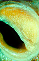
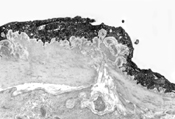
Figure 1. Saphenous vein for bypass surgery
Scanning electron microscope (SEM) image showing a circumferential section through
human saphenous vein with a pedicle of surrounding tissue harvested
for bypass surgery by 'no-touch' technique introduced by Dr. DSR
Souza of Örebro University Hospital, Örebro, Sweden.
See Research page for brief project
description.
Original magnification x35.
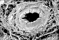
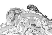
Figure 2. Structure of the vessel wall
Transmission electron microscope (TEM) image demonstrates the main cell types
involved in the build, and hence physiology, of the vascular
wall: this scheme may apply both to arteries and veins. Here,
an example of a coronary vessel (rat) shows vascular endothelium
(intima), vascular smooth muscle (media) and autonomic
perivascular nerves; the latter are located in the surrounding
the vessel tissue (adventitia); Adventitia also consists fibroblasts
together with collagen fibers. Perivascular nerves (axons),
which in this case may be derived from the intra-cardiac ganglia,
convey various types of synaptic vesicles (e.g. small agranular,
and large and small granular vesicles). These vesicles contain
a variety of transmitters/vasoactive agents (depending on the
type of nerve), which when released from the nerve axon varicosity
interact with the relevant receptor on vascular smooth muscle
causing its relaxation or contraction (regulation of the vessel
diameter and hence blood flow). For example, perivascular nerves
deriving from intra-cardiac ganglia (intrinsic cardiac nervous
system), may generate nitric oxide (NO) - a powerful vasorelaxant.
A similar function may apply to vascular endothelium since it
is the source of a variety of vasoactive agents including NO.
Original magnification x8000.
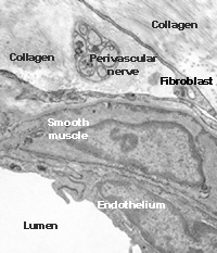
Figure 3: Diseased vascular smooth muscle cell
TEM image of smooth muscle cell of the middle cerebral artery from a Multiple System Atrophy sufferer. Note the accumulation of lipids (dark material) in the cell.
Original magnification x4000
Figure 4: Endothelin 1 (ET-1) in cerebrovascular endothelium
TEM image of human middle cerebral artery immuno-labelled for ET-1 (vasoconstrictor),
demonstrates that the endothelium lining at the artery lumen is
strongly immunoreactive for ET-1 (black stain). Methods:
pre-embedding PAP technique with a polyclonal ET-1 antibody.
Original magnification x10000.
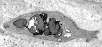
Figure 5: Substance P (SP) in cerebrovascular endothelium
TEM image of human middle cerebral artery immunolabelled for SP
shows one SP-positive (dark-blackish stained) and at
least one SP-negative endothelial cell (lining at the artery
lumen). Methods: pre-embedding PAP technique with a polyclonal
SP antibody.
Original magnification x6000.