


Andrzej Loesch
PhD, DSc. FSB
Professor of Medical Sciences
Visito to Division of Medicine
Royal Free Campus
University College London
Rowland Hill Street
London NW3 2PF, UK
Email: a.loesch@ucl.ac.uk aloesch@talktalk.net
Gallery 4
Perivascular nerves
Click on the image for a large version
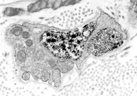
Figure 1: Perivascular autonomic nerve varicosity
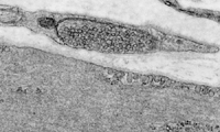
TEM image of an adventitial-medial border (of mouse tibial artery) shows an axon varicosity close to vascular smooth muscle (gap ~130nm); varicosity contains small agranular synaptic vesicles.
Original magnification x30000.
Figure 2: Perivascular axon varicosity containing neuropeptide Y (NPY)
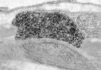
TEM image of a mouse tibial artery shows an NPY-positive axon varicosity (with black immunoprecipitate) close to vascular smooth muscle (gap ~100nm). The varicosity plane contains mostly small agranular synaptic vesicles but one or two large granular vesicles can also be seen. Methods: pre-embedding PAP immunocytochemistry with a polyclonal NPY antibody.
Original magnification x33000
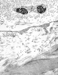
Figure 3: Cerebrovascular nerve fibres (axons) producing NO - 'vasodilator nerves'
TEM image of rat basilar artery demonstrates positive for nNOS (black labels) perivascular axon varicosities close to vascular smooth muscle; varicosities are enclosed by nNOS-negative Schwann cell. Methods: pre-embedding immunogold-silver technique with a polyclonal nNOS antibody.
Original magnification x10000.
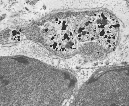
Figure 4: Human cerebrovascular nerves producing NO.
TEM image of human middle cerebral artery immunolabelled for NOS-1 shows two NOS-1-positive axon profiles (with black stain); adjacent cell profiles are NOS-1-negative. NOS-1-positive cerebrovascular axon in the centre of the image contains several labelled large granular vesicles; NOS-1-positive varicosity to the right contains mostly small agranular vesicles but one or two granular vesicles can also be seen. Methods: pre-embedding PAP immunocytochemistry with a polyclonal NOS-1 antibody.
Original magnification x15000.
Figure 5 (A,B): ET-1 in cerebrovascular (vasoconstrictor?) nerves: the origin of the nerves.
LEFT (A) Basilar artery
TEM image of rat basilar artery shows ET-1-positive perivascular axons (black stain) in the adventitia. These axons may originate from the ET-1 positive sensory neurones of trigeminal ganglia and the sympathetic neurones of the superior cervical ganglia. Original mag. x12000.
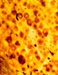
RIGHT (B) Trigeminal ganglion LM image of rat trigeminal ganglion (70 µm vibratome section) shows ET-1-positive neurones (brown stain). Methods: pre-embedding PAP technique with polyclonal ET-1 anti-body. ET-1 positive neurones of sizes 30-40 µm.