


Andrzej Loesch
PhD, DSc, FSB
Professor of Medical Sciences
Visitor to Division of Medicine
Royal Free Campus
University College London
Rowland Hill Street
London NW3 2PF, UK
Email: a.loesch@ucl.ac.uk
aloesch@talktalk.net
Gallery 3
Injured vessels, platelets and leukocytes
Click on the image for a large version
Figure 1: Platelets in injured vessel
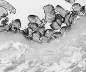
TEM image of injured intima of carotid artery (rat 24h after balloon-angioplasty displays no endothelial cells but clustered platelets at the artery luminal surface. Specimen was stained (black label) with lectin from Bandeirea simplicifolia (BS-I isolectin B4)
Original magnification x5000
Figure 2 (a-d): Leukocyte invasion into vascular wall
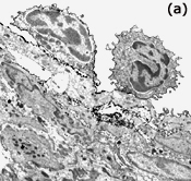
TEM images of injured carotid artery (rat 4-8 weeks after balloon-angioplasty) display various stages of leukocyte migration from the vessel lumen to the neointima. Image (a) shows leukocytes adhered to regenerated endothelium. Images below show: (b) a leukocyte migrating through the endothelial layer, (c) thinned endothelium overlaying a leukocyte, (d) a leukocyte with visible nucleus is located in deeper regions of the neointima. Specimens were stained (black label) with BS-I isolectin B4; label is seen around the leukocyte surface (membrane), some is on endothelial plasmalemma.
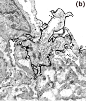
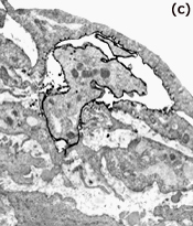
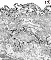
Original magnifications: a x4000, b x8000, c x8000, d x4000.