


Andrzej Loesch
PhD, DSc, FSB
Professor of Medical Sciences
Visitor to Division of Medicine
Royal
Free Campus
University College London
Rowland Hill Street
London NW3 2PF, UK
Email: a.loesch@ucl.ac.uk
aloesch@talktalk.net
Gallery 2
Endothelial vasoactive agents and their receptors
Click on the image for a large version
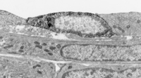
Figure 1: Pulmonary endothelium containing arginine-vasopressin (AVP)
TEM image of rat (neonatal) pulmonary artery showing a profile of AVP-positive endothelial cell; immunoprecipitate (black stain) is associated with extensive granular endoplasmic reticulum. Methods: pre-embedding PAP technique with a polyclonal AVP antibody.
Original magnification x8000.
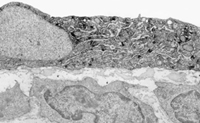
Figure 2: Coronary endothelium containing AVP
TEM image of rat (neonatal) coronary artery showing an AVP-positive endothelial cell; immunoprecipitate (black stain) is seen in the cell cytoplasm. Neighbouring AVP-negative endothelial cell can also be seen. Methods: pre-embedding PAP technique with a polyclonal AVP antibody.
Original magnification x8000.
Figure 3: Myocardial capillary producing NO
TEM image of a myocardial capillary of the mouse heart, immunolabelled for nitric oxide synthase (isoform NOS-3/eNOS), shows that the endothelium lining the inside of the capillary is immunoreactive for NOS-3 (blackish stained)[NOS is the enzyme producing NO]. The capillary is surrounded by myocytes containing a number of mitochondria. Methods:pre-embedding ABC/ExtrAvidin technique with a monoclonal NOS-3 antibody.
Original magnification x8000
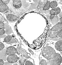
Figure 4: Renal glomerular capillary producing NO
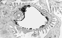
TEM image of a glomerular capillary in the rat kidney, immunolabelled for NOS-3, demonstrates that the endothelium lining the inside of the capillary is immunoreactive for NOS-3 (black stain). The foot processes of the podocytes (immunonegative) embrace the capillary, in this section looking like a corona. Methods:pre-embedding ABC/ExtrAvidin technique with a monoclonal NOS-3 antibody.
Original magnification x6000
Figure 5: P2X6 receptors in human cultured umbilical vein endothelial cells (HUVECs)
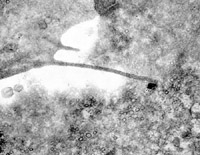
TEM image of HUVECs shows a cellular process of one cell contacting the other cell; at the site of contact, relatively large black label for the P2X6 receptor is seen [Methods: pre-embedding immunogold-silver technique with a polyclonal P2X6 antibody]. Specimen was co-labelled for VE-cadherin [by pre-embedding ABC/ExtrAvidin method with a polyclonal VE-cadherin antibody]; the VE-cadherin labelling (black precipitate) can be seen in cytoplasmic vesicles of the cell to the right. P2X receptors and VE-cadherin are important at junctions formation.
Original magnification x15000.