FLUIDIZATION RESEARCH GROUP
Dr. Paola Lettieri
UCL logo

The Group

Experimental

CFD Modelling

Sustainable Development

X-Ray Imaging

X-ray Imaging

The Fluidization Research Group is home to a unique X-ray Imaging Facility that can give quantitative real time information about the internal flow pattern of a rapidly changing three dimensional system. Over the years, The X-ray facility has been used for both academic and industrial work to study the flow phenomena and fluid-particle interactions involved in dense multiphase systems, to analyse the bubble dynamics, and how the operating conditions influence the reactor performance, efficiency and scale-up.

We are able to analyse X-ray images and provide data that allows quantifying the average fluid-bed voidage, the voidage distribution around bubbles, to carry out the detailed analysis of bubble dynamics and bubble hold-up, together with obtaining expansion and deaeration profiles. X-ray imaging is also used as a means of validation for the CFD models developed within the Group for 
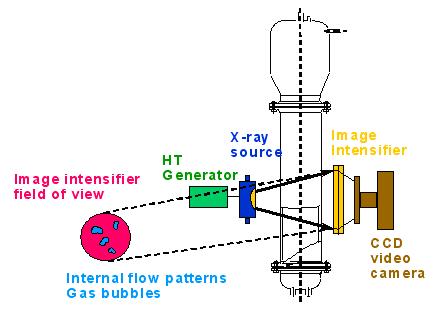
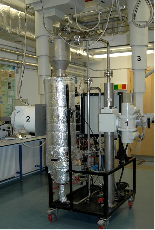
the simulations of multiphase systems.

The Facility

The X-ray equipment is housed in a radiation proof room which contains the X-ray generator, X-ray tube (1) and an image intensifier (2), see Figure below. The X-ray tube and image intensifier are mounted on a twin column ceiling suspension unit, see (3), which allows the columns to be moved along the length of the room. The lateral movement of each column then permits the distance between the tube and image intensifier to be changed. Each of the columns can also be moved in a vertical plane either independently or synchronized as a pair. This motion is motorized and can be remotely
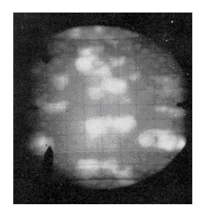
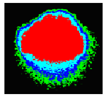
 controlled from outside the room.

The X-ray source is pulsed at 25 fps, where a high energy beam (from 40 to 150 kV) is produced from a rotating anode. The X-ray is designed to operate up to 150 kVp (kilovolts peak). The X-ray pulses are synchronised with an image capturing device and pass through the reactor vessel, where X-ray absorption is proportional to the nature and quantity of material along the path. The X-ray beam emerging from the reactor vessel is amplified using an image intensifier which converts the X-ray absorption patterns into a light image of sufficient brightness and contrast to be recorded by a video camera. As a result, the brightness intensification on the output screen is increased by a factor of 1,000 compared with the brightness of the input screen. The intensifier is optically coupled to 25-75 fps, 1024 x 1024 pixel digital camera. The images from the camera are then sent by fibre optic to a dual processor Industrial PC with redundant (RAID) storage in order to store the real-time image sequence in memory, un-compressed such that no quality is lost. This includes a comprehensive toolkit of image processing functions to allow replay, slow motion, freeze of the images. The image processing functions also allow for image enhancement, sizing and profiles to be extracted. The entire sequence is archived digitally. 

A Schematic and photo of the X-ray Facility and an X-ray image of a single bubble and of a freely bubbling bed are shown below.


X-Ray Schematic

Schematic of the X-ray Imaging Facility at UCL

Freely Bubbling Bed
X-ray Image of a Freely Bubbling Bed

X-ray image of a single bubble
X-ray Rig

Photograph of the X-ray Facility at UCL
rear view


Photograph of the X-ray Facility at UCL
front view

(Created 20 November 2007, last updated 20 February 2009)


University College London - Gower Street - London - WC1E 6BT - Telephone: +44 (0)20 7679 2000 - Copyright © 1999-2007 UCL Disclaimer | Accessibility | Privacy | Advanced Search | Help